Conditions
AC Joint Separation
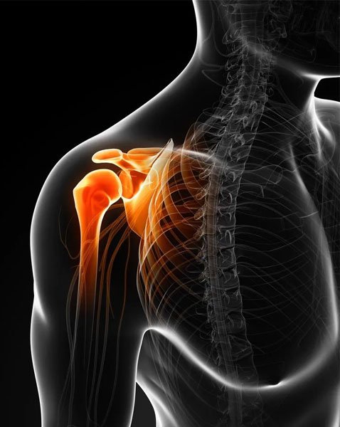
An Acromioclavicular Joint Separation or AC Joint Separation or AC Joint Sprain or simply Shoulder Separation is a common shoulder injury among active people like athletes. In this injury, the clavicle (collar bone) separates from the scapula (shoulder blade). The clavicle and scapula together form the socket that holds the ball of the upper arm bone. AC Joint Separation is generally caused due to a direct injury like a fall or a blow to that spot. It is a common sports injury, frequently seen in football and hockey players and cyclists.
Classification of AC Joint Injuries
AC Joint injuries range from very mild (Grade 1) to a severe injury (Grade 6). AC Joint Dislocations are classified by Tossy (3-grade classification), Allman (3-grade classification) and Rockwood (6-grade classification). Rockwood’s classification is the one most commonly used.
Symptoms of AC Joint Separation
Symptoms include pain and swelling on the shoulder. The pain generally increases when trying to make overhead movements or trying to sleep on the affected side. In some cases, there will be limited movement in the shoulder area and in some severe cases, a lump will be formed on top of the shoulder joint.
Diagnosis of Shoulder Separation
Your doctor might diagnose AC joint separation by any or all of the following methods: Medical history, Physical examination, X-ray or some tests to evaluate the pain and the range of motion.
Treatment of Acromioclavicular Joint Separation
Most AC Joint injuries don’t need surgery however in some severe cases surgery is required. People with low grade AC joint injuries will be put on standard medical treatment which includes:
- Rest
- Icing
- Using Sling
- Anti-inflammatory Drugs
- Physical Therapy
Such injuries (generally Grade 1 to 3) will be healed in two to three weeks.
People with high grade AC Joint injuries (generally Grade 4 to 6 and in some cases Grade 3) will be required to undergo surgery in cases where standard non-surgical methods does not help relieve the pain and swelling. A surgeon will try to put the clavicle back to scapula. For this, a variety of implants are available as per the need of the patient. These surgeries are generally performed using Arthroscopy, which is less painful and with better recovery time. However, in some cases open surgical methods are used. Suitable rehabilitation will be required post-surgery including wearing of sling, medications and physio/occupational therapy.
Causes of AC joint separation:
More often than not, AC joint separation occurs out of a fall directly onto the shoulder. As a result, the ligaments that surround and stabilize the AC joint get injured. If the fall is a severe, the ligaments that support the underside of the clavicle are also torn, resulting in the separation of the scapula and the collar bone. The fall results in the shoulder blade moving down due to the weight of the arm, thus creating a bump or bulge at the top of the shoulder.
The AC joint separation injury can range from a mild injury without any further complications or a complete tear with a large bulge. With proper treatment and rehabilitation, good pain-free range of motion of the shoulder is possible even with a very large bulge. A severe deformity may require a longer time for the pain-free motion to return. If the AC joint separation is mild, there may be a sprain of the AC joint ligament that does not affect the collar bone and it looks normal on imaging such as x-ray. However, a serious injury tears the AC joint, either sprains or slightly tears the coracoclavicular ligament. It may also cause dislocation of the collar bone with a small bulge. In the case of a complete AC joint separation, acromioclavicular joint and coracoclavicular ligaments are torn extensively. This puts the acromioclavicular ligament completely out of alignment and there will be a larger bulge.
Identifying an AC joint separation:
If there is AC joint deformity, the injury can be identified easily. However, if the intensity of the injury is less, the location of the tenderness and x-rays can help the doctor to arrive at a diagnosis. At times, carrying a weight in hand makes the injury more prominent, thereby making the injury more significant on x-rays.